Useful things to know.

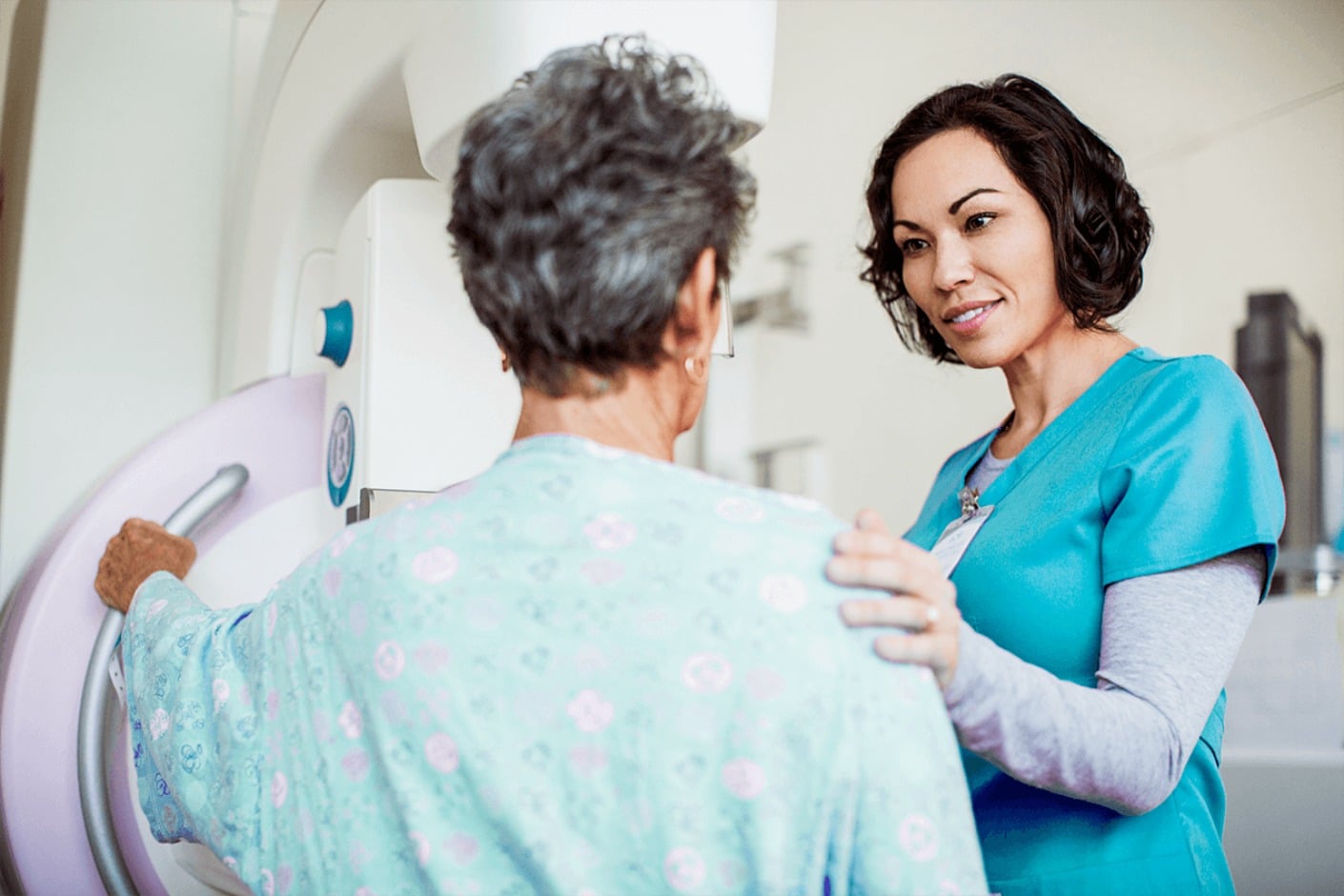
During the exam:
Does it hurt?
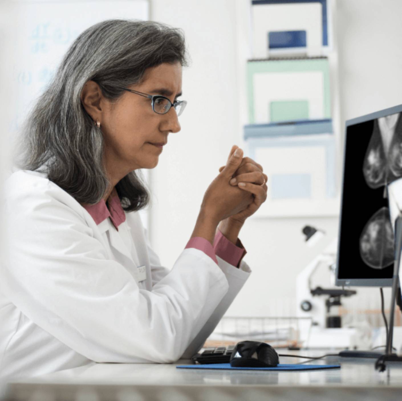
After your exam:

During the exam:

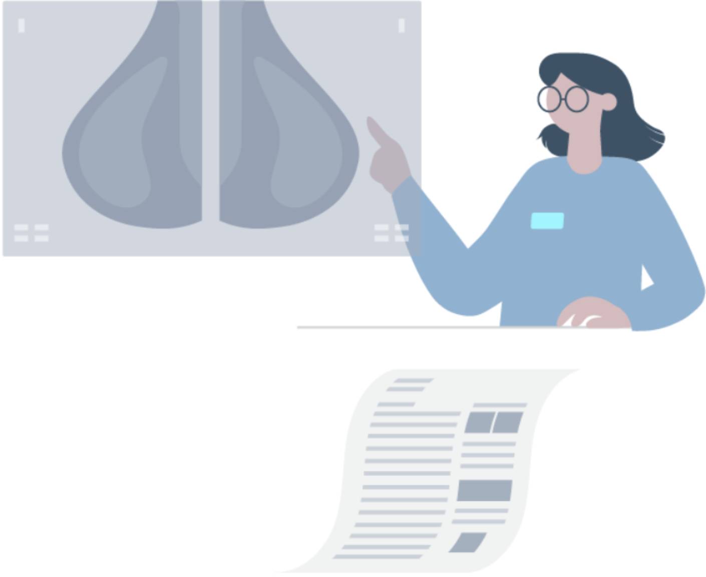
More tests may be needed after your mammogram for a number of reasons:
It's important to advocate for your own health.
Empower yourself with information from these breast care resources.
1. Pisano et al. Diagnostic Performance of Digital versus Film Mammography for Breast–Cancer Screening. NEJM 2005;353:1773.
Disclaimer: The content on this site is for informational purposes only. The content is not intended to be medical advice, diagnosis, or treatment, or a substitute for such advice, diagnosis, or treatment. You should always consult with your healthcare provider for medical advice, diagnosis, and treatment, including your specific medical needs. Lunit does not recommend or endorse any specific methods of supplemental screening or treatment.